
Welcome to the David Lab Website!
The David Lab at UC Davis utilizes numerous tools of chemical biology to explore the complex mechanistic details of DNA repair enzymes. DNA repair proteins such as MutY and NEIL, among the targets of research of the David Lab, help catalyze necessary repair of oxidative DNA damage, and are critical to maintaining genomic integrity in organisms living in the oxygen-rich environment of Earth. The David Lab is headed by Dr. Sheila S. David, who has led at the forefront of DNA repair research for the last 30 years. The David Lab continues to push forward the world’s current understanding of DNA repair enzymes by leveraging our unique expertise in DNA repair enzymology while venturing into new areas and collaborating with scientists around the world.
Featured Articles:
Structure of Human MUTYH and Functional Profiling of Cancer-Associated Variants Reveal an Allosteric Network Between its [4Fe-4S] Cluster Cofactor and Active Site Required for DNA Repair
Nature Communications
“MUTYH is a clinically important DNA glycosylase that thwarts mutations by initiating base-excision repair at 8-oxoguanine (OG):A lesions. The roles for its [4Fe-4S] cofactor in DNA repair remain enigmatic. Functional profiling of cancer-associated variants near the [4Fe-4S] cofactor reveals that most variations abrogate both retention of the cofactor and enzyme activity. Surprisingly, R241Q and N238S retained the metal cluster and bound substrate DNA tightly, but were completely inactive. We determine the crystal structure of human MUTYH bound to a transition state mimic and this shows that Arg241 and Asn238 build an H-bond network connecting the [4Fe-4S] cluster to the catalytic Asp236 that mediates base excision. The structure of the bacterial MutY variant R149Q, along with molecular dynamics simulations of the human enzyme, support a model in which the cofactor functions to position and activate the catalytic Asp. These results suggest that allosteric cross-talk between the DNA binding [4Fe-4S] cofactor and the base excision site of MUTYH regulate its DNA repair function.“
David Lab Authors:
Dr. Carlos Trasviña-Arenas, Mohammad Hashemian, Merve Demir, Prof. Sheila S. David
Crystal Structure of MutYX: A Novel Clusterless Adenine DNA Glycosylase with a Distinct C-terminal Domain and 8-Oxoguanine Recognition Sphere
bioRxiv
“The [4Fe-4S] cluster is an important cofactor of the base excision repair (BER) adenine DNA glycosylase MutY to prevent mutations associated with 8-oxoguanine (OG). Several MutYs lacking the [4Fe-4S] cofactor have been identified. Phylogenetic analysis shows that clusterless MutYs are distributed in two clades suggesting cofactor loss in two independent evolutionary events. Herein, we determined the first crystal structure of a clusterless MutY complexed with DNA. On the basis of the dramatic structural divergence from canonical MutYs, we refer to this as representative of a clusterless MutY subgroup “MutYX”. Interestingly, MutYX compensates for the missing [4Fe-4S] cofactor to maintain positioning of catalytic residues by expanding a pre-existing α-helix and acquisition of the new α-helix. Surprisingly, MutYX also acquired a new C-terminal domain that uniquely recognizes OG using residue Gln201 and Arg209. Adenine glycosylase assays and binding affinity measurements indicate that Arg209 is the primary residue responsible to specificity for OG:A lesions, while Gln201 bridges OG and Arg209. Surprisingly, replacement of Arg209 and Gln201 with Ala increases activity toward G:A mismatches. The MutYX structure serves as an example of devolution, capturing structural features required to retain function in the absence of a metal cofactor considered indispensable.“
David Lab Authors:
Dr. Carlos Trasviña-Arenas, Mohammad Hashemian, Melody Malek, Steven Merrill, Prof. Sheila S. David
FSHing for DNA Damage: Key Features of MutY Detection of 8-Oxoguanine:Adenine Mismatches
Accounts of Chemical Research

“Base excision repair (BER) enzymes are genomic superheroes that stealthily and accurately identify and remove chemically modified DNA bases. DNA base modifications erode the informational content of DNA and underlie many disease phenotypes, most conspicuously, cancer. The “OG” of oxidative base damage, 8-oxo-7,8-dihydroguanine (OG), is particularly insidious due to its miscoding ability that leads to the formation of rare, pro-mutagenic OG:A mismatches. Thwarting mutagenesis relies on the capture of OG:A mismatches prior to DNA replication and removal of the mis-inserted adenine by MutY glycosylases to initiate BER. The threat of OG and the importance of its repair are underscored by the association between inherited dysfunctional variants of the MutY human homologue (MUTYH) and colorectal cancer, known as MUTYH-associated polyposis (MAP). Our functional studies of the two founder MUTYH variants revealed that both have compromised activity and a reduced affinity for OG:A mismatches. Indeed, these studies underscored the challenge of the recognition of OG:A mismatches that are only subtly structurally different than T:A base pairs. Since the original discovery of MAP, many MUTYH variants have been reported, with most considered to be “variants of uncertain significance.” To reveal features associated with damage recognition and adenine excision by MutY and MUTYH, we have developed a multipronged chemical biology approach combining enzyme kinetics, X-ray crystallography, single-molecule visualization, and cellular repair assays. In this review, we highlight recent work in our laboratory where we defined MutY structure–activity relationship (SAR) studies using synthetic analogs of OG and A in cellular and in vitro assays. Our studies revealed the 2-amino group of OG as the key distinguishing feature of OG:A mismatches. Indeed, the unique position of the 2-amino group in the major groove of OGsyn:Aantimismatches provides a means for its rapid detection among a large excess of highly abundant and structurally similar canonical base pairs. Furthermore, site-directed mutagenesis and structural analysis showed that a conserved C-terminal domain β-hairpin “FSH’’ loop is critical for OG recognition with the “His” serving as the lesion detector. Notably, MUTYH variants located within and near the FSH loop have been associated with different forms of cancer. Uncovering the role(s) of this loop in lesion recognition provided a detailed understanding of the search and repair process of MutY. Such insights are also useful to identify mutational hotspots and pathogenic variants, which may improve the ability of physicians to diagnose the likelihood of disease onset and prognosis. The critical importance of the “FSH” loop in lesion detection suggests that it may serve as a unique locus for targeting probes or inhibitors of MutY/MUTYH to provide new chemical biology tools and avenues for therapeutic development.“
David Lab Authors:
Chandrima Majumdar, Merve Demir, Steven Merrill, Mohammad Hashemian, Prof. Sheila S. David
For the latest David Lab updates, check out the News section.
Click on Research for an overview of The David Lab’s research.
A thorough list of publications is available in the Publications section.
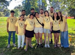
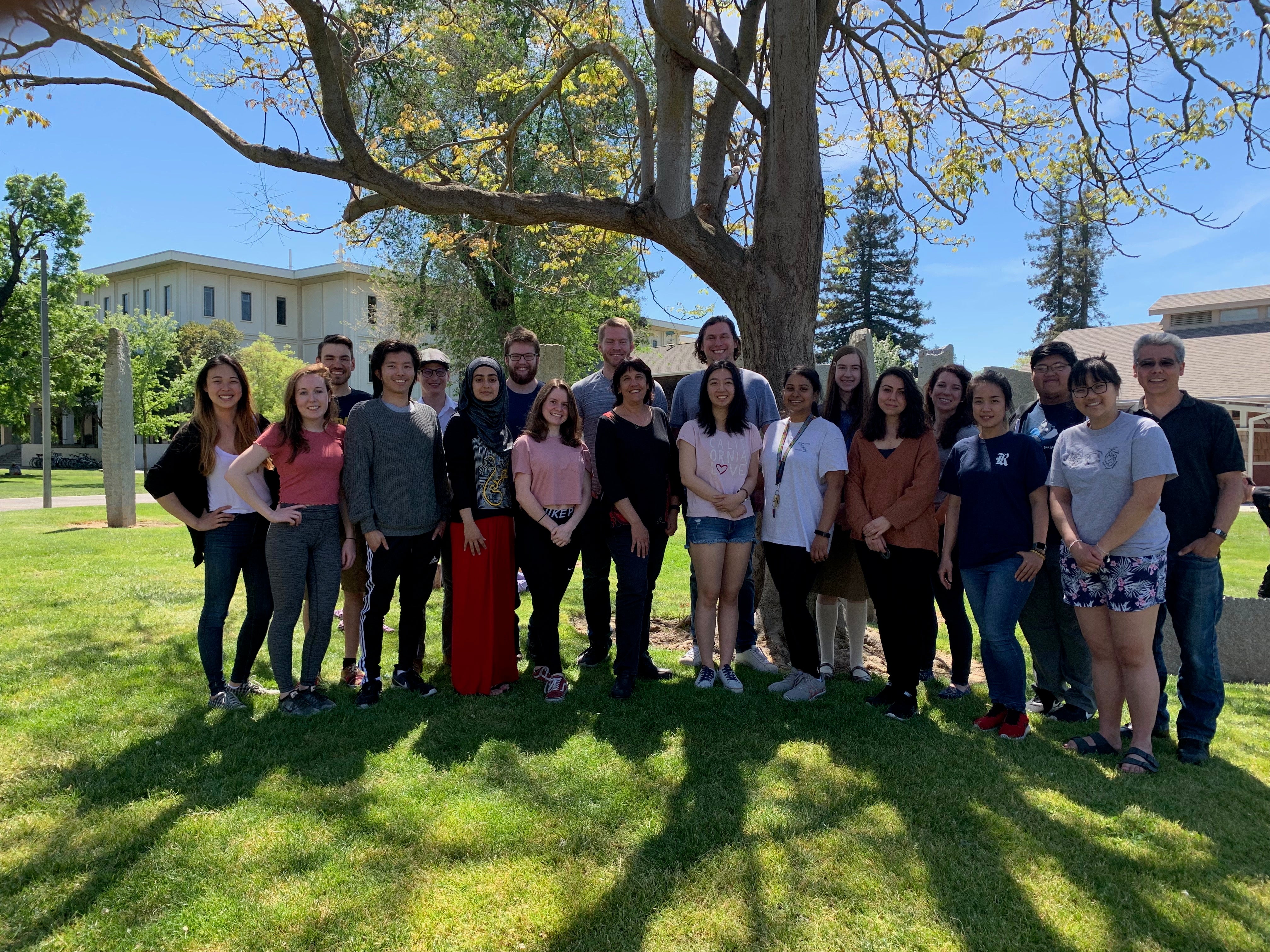
David Lab Members
Keywords: #DavidLab #TheDavidLab #UCDavis #DNA #DNARepair #Muty #Mutyh #8OG #enzymes #ModifiedOligonucleotides #ModifiedNucleosides #OrganicSynthesis #Synthesis #BaseExcisionRepair #BER #NEIL #ChemicalBiology #Chemistry #SheilaDavid #UCDavisChemistry #glycosylase #DNARepairUCDavis